About Embryo Library

The Embryo Library is the biggest worldwide digital atlas of time‑lapse embryo images. Each of the thousands time‑lapse embryo images features monitored morphokinetic markers.
Time‑lapse embryo images were recorded by partner clinics. All pictures were anonymized and evaluated by CATI’s artificial intelligence (AI) and then carefully reviewed by senior embryologists to make this embryo collection a high-quality educative material.
Provided Images
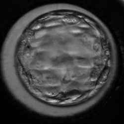
All images were obtained by EmbryoScope ® in 30-minute interval. A time‑lapse embryo image is a 72-DPI image measuring 250px width, 250px height.
The Embryo Evaluation Process
Automated time‑lapse embryo evaluation goes through three phases – Artificial Intelligence (AI) detection, applying rules and manual review. Only after undergoing these stages, each embryo is qualified for publishing in the collection.
AI Embryo Detection
CATI AI is responsible for detecting the embryo in a time‑lapse image. It detects morphokinetic markers that include area of embryo, cell cleavage timing, blastocoel expansion, blastocoel collapse timing, and the activity of cells.
Applying Grading Model Rules
The senior embryologists behind CATI developed a smart day‑five embryos grading system which defines rules based on the timing of morphokinetic markers. These rules are applied to data detected from the previous phase.
Manual Review
To make sure that each and every embryo is correctly evaluated and annotated, our senior embryologists carefully observe all embryos and verify machine-detected annotations.